

2 Trauma Nursing Research Center, Kashan University of Medical Sciences, Kashan, Iran
3 Department of Epidemiology, Kashan University of Medical Sciences, Kashan, Iran
4 Obstetrician and Gynecologist, Kashan, Iran
*Corresponding author: Masoumeh Abedzadeh-Kalahroudi, Trauma Nursing Research Center, Kashan University of Medical Sciences, Kashan, Iran. Email: abedzadeh@kaums.ac.ir
EXTENDED ABSTRACT
Background
Preterm birth, defined as birth before 37 completed weeks of gestation, remains a major cause of neonatal mortality and morbidity. It is reported in approximately 7-12% of pregnancies in developed countries and may be more frequent where access to neonatal intensive care is limited [1, 2]. In Iran, the incidence of preterm birth increased from 8.9% to 12.1% between 2005 and 2019 [3]. Several pharmacologic approaches are used to prevent or delay preterm delivery, and progestogens have become an important option in women at increased risk [4, 5]. Proposed mechanisms of progesterone include anti-inflammatory activity, inhibition of myometrial gap-junction formation, effects on cervical ripening, modulation of potassium channels, inhibition of oxytocin and prostaglandin pathways, and vasodilatory effects on the uterine circulation [6-10]. Natural progesterone can be administered orally or vaginally, whereas hydroxyprogesterone caproate is administered by injection [11]. Uncertainty persists regarding the optimal preparation, route, dose, and timing of progesterone therapy [12, 13]. Micronized progesterone (Utrogestan) was developed to improve oral absorption and has been used during pregnancy, while 17-alpha-hydroxyprogesterone caproate (Proloton) is an injectable preparation requiring intermittent intramuscular administration [15-18]. Previous Doppler studies have reported both vasodilatory effects and little or no change in maternal or fetal circulation after progesterone therapy [8, 9, 19-24]. Therefore, this randomized trial compared oral micronized progesterone with intramuscular hydroxyprogesterone caproate in women at risk of preterm labor, focusing on Doppler indices of the uterine, umbilical, and fetal middle cerebral arteries.
Methods
This randomized, single-blind clinical trial included 80 pregnant women at risk of preterm labor who were referred to Shabihkhani and Shahid Beheshti hospitals in Kashan during Iranian calendar year 1400. The sample size was 40 women per group. Risk of preterm labor was defined by a history of preterm birth, a second-trimester cervical length below 25 mm, or regular uterine contractions requiring hospitalization. Eligible women had a singleton pregnancy at 20-34 weeks of gestation confirmed by last menstrual period and ultrasonography, cervical length below 25 mm on transvaginal ultrasonography, intact membranes, a history of preterm delivery or regular uterine contractions, cervical dilatation of at least 2 cm, and no hypertension. Exclusion criteria were failure to return for follow-up Doppler ultrasonography, incorrect medication use, known fetal anomaly or fetal death, treatment with tocolytics, progesterone hypersensitivity, or an indication for immediate delivery such as fetal distress or severe bleeding. After written informed consent, uterine, umbilical, and fetal middle cerebral artery Doppler ultrasonography was performed by a perinatologist blinded to treatment allocation. Randomization used computer-generated block allocation with block sizes of four and six and sequential opaque envelopes. Women assigned to Utrogestan received oral micronized progesterone 200 mg twice daily for 48 hours; those assigned to Proloton received 500 mg hydroxyprogesterone caproate intramuscularly. Doppler ultrasonography was repeated one week later by an examiner unaware of the intervention. Examinations were performed with a Medison 200 ultrasound system and a 2-5 MHz abdominal transducer. Pulsatility index (PI), resistance index (RI), peak systolic velocity (PSV), and systolic-to-diastolic ratio (S/D) were measured in the uterine, umbilical, and fetal middle cerebral arteries. Data were analyzed using SPSS version 16. Within-group comparisons used the Wilcoxon signed-rank test; between-group comparisons used Fisher's exact test, independent-samples t test, or Mann-Whitney test as appropriate. ANCOVA was used to adjust for baseline differences in Doppler indices, and linear regression evaluated predictors of post-treatment values. Statistical significance was defined as P<0.05. The study was approved by the Kashan University of Medical Sciences ethics committee (IR.KAUMS.MEDNT.REC.1398.096; grant 98146), and the trial was registered as IRCT20200201046324N1.
Results
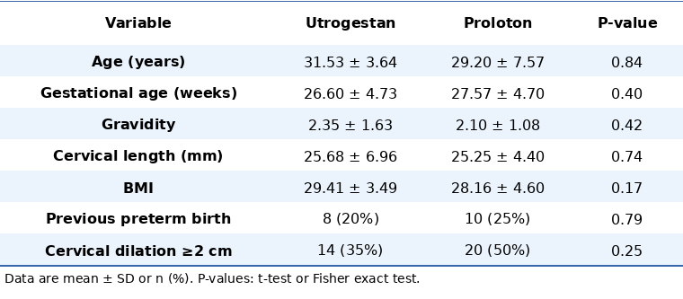
The two treatment groups were comparable at baseline with respect to maternal age, body mass index, gestational age, gravidity, history of preterm delivery, cervical length, and initial cervical dilatation. Mean maternal age was 31.53±3.64 years in the Utrogestan group and 29.20±7.57 years in the Proloton group (P=0.84). Mean gestational age was 26.60±4.73 and 27.57±4.70 weeks, respectively (P=0.40). Previous preterm birth was reported in 8 women (20%) receiving Utrogestan and 10 (25%) receiving Proloton (P=0.79), while cervical dilatation of at least 2 cm was present in 14 (35%) and 20 (50%), respectively (P=0.25).
Table 1. Demographic and reproductive characteristics of patients in the two study groups.
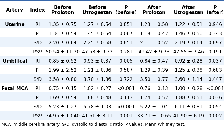
Within-group Doppler analysis showed that uterine artery RI, PI, and S/D decreased significantly after Proloton, with P=0.030, P<0.001, and P=0.019, respectively. In the Utrogestan group, uterine artery RI and S/D decreased significantly (P<0.001 and P=0.002), whereas the change in PI was not significant (P=0.085). Umbilical artery S/D decreased from 3.58±0.80 to 3.50±0.77 after Proloton (P=0.021), while the reduction from 3.70±1.36 to 3.60±1.14 after Utrogestan was not significant (P=0.090). For the fetal middle cerebral artery, Utrogestan was associated with a significant change in S/D from 5.78±1.03 to 6.11±0.81 (P<0.001); other within-group changes in this artery were not statistically significant.
Before treatment, several Doppler indices differed between groups, including umbilical artery RI and fetal middle cerebral artery RI, S/D, and PSV. After treatment, unadjusted between-group comparisons continued to show differences in umbilical artery RI (P=0.037), fetal middle cerebral artery RI (P<0.001), PI (P=0.036), and PSV (P=0.002), while the difference in fetal middle cerebral artery S/D was borderline (P=0.054). Because baseline values of some Doppler indices differed between groups, ANCOVA was used to control for these potential confounders. After adjustment, only fetal middle cerebral artery S/D and PSV remained significantly different between the two treatment groups (P<0.05). The article concluded that the two progesterone preparations had generally similar effects on uterine and umbilical circulation, whereas Proloton produced the more favorable effect on fetal cerebral circulation.
Table 3. Comparison of Doppler indices of the uterine, umbilical, and fetal middle cerebral arteries before and after drug administration between the two groups.
Linear regression further showed that post-treatment Doppler values were influenced not only by treatment assignment but also by maternal age, gestational age, previous preterm birth, BMI, and the corresponding pre-intervention PI, RI, PSV, or S/D values. No adverse effects were reported with either Utrogestan or Proloton during the study.
Conclusion
Oral micronized progesterone and intramuscular hydroxyprogesterone caproate produced broadly similar effects on uterine and umbilical blood flow in women at risk of preterm labor. After adjustment for baseline Doppler differences, the principal between-group differences involved fetal middle cerebral artery S/D and PSV, favoring Proloton. On the basis of the study findings, either preparation may be selected according to patient preference and available facilities, while Proloton may provide a greater effect on fetal cerebral circulation.
Keywords: Hydroxyprogesterone Caproate, Maternal and Fetal Blood Circulation, Micronized Progesterone, Preterm Labor
REFERENCES
- Cunningham F, Leveno K, Bloom S, Dashe JS, Spong C, Hauth J, et al. Williams Obstetrics: 25th Edition. McGraw-Hill Education. 2018.
- De Tejada BM, Karolinski A, Ocampo M, Laterra C, Hösli I, Fernández D, et al. Prevention of preterm delivery with vaginal progesterone in women with preterm labour (4P): randomised double-blind placebo-controlled trial. BJOG: An International Journal of Obstetrics & Gynaecology. 2015;122(1):80-91. DOI: 10.1111/1471-0528.13061
- Abbassinia H, Dashti S, Gholami R, Ghalekhondabi L, Borumandnia N. Increasing Trend and Influencing Factors of Pre-term Labor in Iran from 2005 to 2019. Iran J Pediatr. 2023;33(6):e139823. DOI: 10.5812/ijp-139823
- Breuking SH, De Ruigh AA, Hermans FJR, Schuit E, Combs CA, de Tejada BM, et al. Progestogen maintenance therapy for prolongation of pregnancy after an episode of preterm labour: A systematic review and meta-analysis. BJOG: An International Journal of Obstetrics & Gynaecology. 2023;130(11):1306-16. DOI: 10.1111/1471-0528.17499
- Haghighi L, Rashidi M, Najmi Z, Homam H, Hashemi N, Mobasseri A, et al. Comparison of intramuscular progesterone with oral nifedipine for treating threatened preterm labor: A randomized controlled trial. Med J Islam Repub Iran. 2017;31:56. PMID: 29445685 DOI: 10.14196/mjiri.31.56
- Romero R. Prevention of spontaneous preterm birth: the role of sonographic cervical length in identifying patients who may benefit from progesterone treatment. Ultrasound Obstet Gynecol. 2007;30(5):675-86. DOI: 10.1002/uog.5174
- Da Fonseca EB, Bittar RE, Carvalho MH, Zugaib M. Prophylactic administration of progesterone by vaginal suppository to reduce the incidence of spontaneous preterm birth in women at increased risk: a randomized placebo-controlled double-blind study. Am J Obstet Gynecol. 2003;188(2):419-24. PMID: 12592250 DOI: 10.1067/mob.2003.41
- Abd El Hameed AA. Vaginal versus intramuscular progesterone in the prevention of preterm labor and their effect on uterine and fetal blood flow. Middle East Fertility Society Journal. 2012;17(3):163-9. DOI: 10.1016/j.mefs.2011.12.003
- Czajkowski K, Sienko J, Mogilinski M, Bros M, Szczecina R, Czajkowska A. Uteroplacental circulation in early pregnancy complicated by threatened abortion supplemented with vaginal micronized progesterone or oral dydrogesterone. Fertil Steril. 2007;87(3):613-8. PMID: 17126337 DOI: 10.1016/j.fertnstert.2006.07.1506
- Deichert U, Albrand-Thielmann C, Van de Sandt M. Doppler-sonographic pelvic blood flow measurements and their prognostic value in terms of luteal phase and implantation: The clinical value of Doppler ultrasound. Human Reproduction. 1996;11(8):1591-3. DOI: 10.1093/oxfordjournals.humrep.a019449
- Kuon RJ, Shi SQ, Maul H, Sohn C, Balducci J, Maner WL, et al. Pharmacologic actions of progestins to inhibit cervical ripening and prevent delivery depend on their properties, the route of administration, and the vehicle. Am J Obstet Gynecol. 2010;202(5):455.e1-9. DOI: 10.1016/j.ajog.2010.03.025
- Ibrahim M, Ramy ARM, Younis MA-F. Progesterone supplementation for prevention of preterm labor: a randomized controlled trial. Middle East Fertility Society Journal. 2010;15(1):39-41. DOI: 10.1016/j.mefs.2010.03.007
- Meis PJ, Goldenberg RL, Mercer BM, Iams JD, Moawad AH, Miodovnik M, et al. The preterm prediction study: risk factors for indicated preterm births. Am J Obstet Gynecol. 1998;178(3):562-7. PMID: 9539527 DOI: 10.1016/s0002-9378(98)70439-9
- Akbari S, BM, Mohtasham N. Evaluation of the effect of progesterone on prevention of preterm delivery and its complications. SJKU. 2009;14(3):11-9.
- Marinov B, Petkova S, Dukovski A, Georgiev G, Garnizov T, Manchev V, et al. Utrogestan and high risk pregnancy. Akusherstvo i ginekologiia. 2004;43(5):22-4.
- Zhu X, Zhang X, Fu Y. Utrogestan as an effective oral alternative for preventing premature luteinizing hormone surges in women undergoing controlled ovarian hyperstimulation for in vitro fertilization. Medicine. 2015;94(21):e909. PMID: 26020402 DOI: 10.1097/MD.0000000000000909
- Navathe R, Berghella V. Progesterone as a tocolytic agent for preterm labor: a systematic review. Curr Opin Obstet Gynecol. 2016;28(6):464-9. DOI: 10.1097/GCO.0000000000000327
- Hydroxyprogesterone (Injection Route) Side Effects. 2007. [Link]
- Agra IK, Brizot ML, Miyadahira MY, Carvalho MH, Francisco RP, Zugaib M. The effect of prenatally administered vaginal progesterone on uterine artery Doppler in asymptomatic twin pregnancies. Eur J Obstet Gynecol Reprod Biol. 2016;205:11-4. DOI: 10.1016/j.ejogrb.2016.08.016
- Barda G, Ben-Haroush A, Barkat J, Malinger G, Luria O, Golan A, et al. Effect of vaginal progesterone, administered to prevent preterm birth, on impedance to blood flow in fetal and uterine circulation. Ultrasound Obstet Gynecol. 2010;36(6):743-8. PMID: 20196070 DOI: 10.1002/uog.7606
- Borna S, Borna H, Gotbizadeh F, Jahani M. Evaluation of Progesterone Effects on Fetal Doppler Velocimetry. JOGCR. 2016;1(3). DOI: 10.17795/ojcr-9399
- Vafaei H, Zamanpour T, Shahraki HR. Preterm birth prevention: effects of vaginal progesterone administration on blood flow impedance in uterine-fetal circulation by Doppler sonography. Glob J Health Sci. 2015;8(7):172-8. PMID: 26925899 DOI: 10.5539/gjhs.v8n7p172
- Maged AM, Shoab AY, Hussein EA, Alsawaf AH, Mahmoud DS, AbdAllah AA, et al. The Effect of Antenatal Vaginal Progesterone Administration on Uterine, Umbilical, and Fetal Middle Cerebral Artery Doppler Flow: A Cohort Study. Am J Perinatol. 2020;37(5):491-6. DOI: 10.1055/s-0042-1756680
- Niromanesh S, Farzin Moghadam S, Rahimi Sherbaf F. Maternal and Fetal Doppler Blood Flow Velocimetry Changes in the Management of Asymptomatic Preterm Labor With Vaginal Progesterone Tablet. Acta Med Iran. 2018;56(6):398-404.
- Hoffman GE, Merchenthaler I, Zup SL. Neuroprotection by ovarian hormones in animal models of neurological disease. Endocr. 2006;29(2):217-31. DOI: 10.1385/ENDO:29:2:217
- Wagner CK. Progesterone receptors and neural development: a gap between bench and bedside? Endocrinology. 2008;149(6):2743-9. DOI: 10.1210/en.2008-0049